


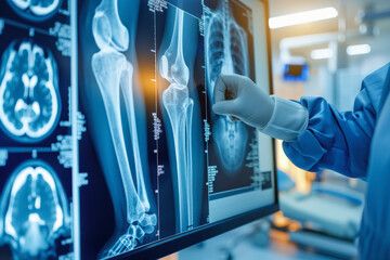
GLP-1 and Bone Density: What Rapid Weight Loss Does to Your Skeleton


EllieMD
5 min read
Significant weight loss can reduce bone density, and GLP-1 therapy is no exception. Here is what the research shows, who is most at risk, and what actually protects bone during weight loss.
Bone density is not a topic that comes up in most GLP-1 program discussions, and for the majority of patients it does not need to be a primary concern. But for specific groups, particularly postmenopausal women, older adults over 65, people with a family history of osteoporosis, and those who lose weight very rapidly, bone density during GLP-1 therapy is worth understanding and monitoring.
Why Weight Loss Affects Bone
Bone is living tissue that responds to mechanical load. The stress of body weight on the skeleton is one of the primary stimuli for maintaining bone density. When bones bear weight regularly, osteoblasts, the cells that build new bone, remain active. When mechanical load decreases, as it does when body weight falls, the stimulus for bone formation decreases alongside it.
This is a well-established relationship. Astronauts experience significant bone loss during weightlessness for the same mechanical reason. Patients who lose weight through bariatric surgery, which produces the most rapid and dramatic weight loss available, show measurable bone density reduction at weight-bearing sites in the years following surgery.
GLP-1 therapy produces weight loss at a slower rate than bariatric surgery, which generally means the bone effects are more modest. But for patients losing 15 to 20 percent of body weight over one to two years, measurable changes in bone mineral density at the hip and spine have been documented in some studies.
The hormonal dimension adds another layer. Adipose tissue is a source of estrogen through aromatization of androgens. Women with obesity often have higher estrogen levels from this peripheral production than lean women. As weight falls significantly, this peripheral estrogen source diminishes, which can accelerate bone loss in women who are already at risk from postmenopausal estrogen decline.
The GLP-1 Receptor on Bone: A Complicating Factor
GLP-1 receptors are expressed on osteoblasts and osteoclasts, the cells responsible for bone formation and resorption respectively. Animal studies have generally shown that direct GLP-1 receptor activation has bone-protective effects, promoting osteoblast activity and inhibiting osteoclast function. This suggests that GLP-1 medications may have a partially protective direct effect on bone that partially counteracts the mechanical unloading effect of weight loss.
Human data on this question is mixed and does not yet clearly resolve whether GLP-1 receptor agonists are net bone-protective, bone-neutral, or mildly bone-reductive over typical treatment periods. The honest clinical position is that the direct receptor effects appear potentially beneficial but the weight loss effect on bone is real and should be managed appropriately for high-risk patients.
Who Should Pay Particular Attention
Postmenopausal women are the group where bone density during GLP-1 therapy warrants the most clinical attention. The combination of existing estrogen-related bone loss, the loss of peripheral estrogen from reduced adipose tissue, and the mechanical unloading from weight loss creates a confluence of factors that can accelerate bone loss in women who are already at elevated fracture risk.
Adults over 65 of either sex have lower baseline bone density and reduced bone remodeling reserve, making weight loss-related bone changes more clinically significant.
Patients with a personal or family history of osteoporosis, or those who have already had a fragility fracture, should discuss bone monitoring specifically with their physician before and during significant weight loss.
What Actually Protects Bone During Weight Loss
Resistance training is the most evidence-based intervention for maintaining bone density during weight loss. The mechanical loading of resistance exercise directly stimulates osteoblast activity and bone mineral deposition in the specific bones loaded. Weight-bearing resistance exercise, squats, lunges, deadlifts, and similar movements, loads the hip and spine, the sites most relevant to fracture risk in aging adults.
Calcium and vitamin D adequacy are foundational. Bone cannot be maintained without adequate substrate. Calcium requirements increase during periods of significant dietary restriction, and vitamin D is required for calcium absorption. Your physician can assess adequacy through serum 25-hydroxyvitamin D and dietary history.
For postmenopausal women on GLP-1 therapy with elevated fracture risk, a baseline DEXA scan before starting treatment provides a documented starting point and allows meaningful monitoring of change over the treatment period. A follow-up scan at 12 to 18 months gives concrete data on whether bone density is being maintained.
Learn more at the EllieMD YouTube Channel.
Individual results may vary. All prescriptions require approval by a licensed medical provider. Compounded medications are not FDA-approved. EllieMD facilitates access to independent healthcare providers and pharmacies and does not provide medical care or dispense medications.
Related Articles
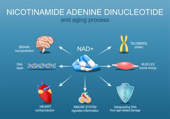
NAD+ Oral Drops with GLP-1: What the Combined Formulation Is Designed to Do
EllieMD
GLP-1 and Joint Pain: The Connection Between Weight, Inflammation, and Chronic Pain
EllieMD
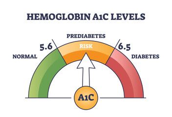
What Prediabetes Actually Means and Why GLP-1 Therapy Is Relevant
EllieMD

GLP-1 Side Effects: What to Expect and How to Manage Them
EllieMD

GLP-1 and Testosterone: What Men Need to Know About the Hormonal Connection
EllieMD
How Does GLP-1 Work in the Body? A Plain-Language Explanation
EllieMD
Get the latest updates and exclusive offers by subscribing to our newsletter.
© 2026 EllieMD LLC. All rights reserved.
